Microscope structure
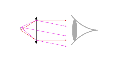
As a tool to visually observe small objects located very near the optical system, a microscope must transform a real object located at a finite distance of the system into a virtual image, located at infinity for a perfect eye, and with a angular size larger than the one seen by the naked eye when the object is placed at the punctum proximum the simplest system verifying those criteria is represented on figure 1. It is composed of a unique thin lens whose focal plane coincides with the observed object. With this configuration, the magnification is larger than \(1\) if the lens focal length is shorter than the distance object-eye when the object is located at the punctum proximum, and, in fact, the shorter the focal lens and the larger the magnification. The magnifying glass works on this principle.
This system can be improved by replacing the simple lens by a singlet or a doublet with aspherical surfaces. But even with this improvement, it will exhibit strong field limitations due to the field aberrations that can't be corrected, and to the vignetting introduced by the eye pupil which is the system field diaphragm. In practice, the magnification and numerical aperture that can be used remain rather small.
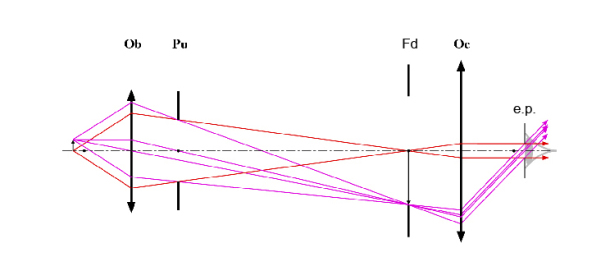
One solution to those problems is shown on figure 2. This compound system, composed of an objective[1] and an ocular[2], allows resolving the problem of pupils conjugations (the eye pupil vignetting is suppressed, since the eye pupil can be placed at the exit pupil of the microscope), and of field aberrations (the full system is composed of many lenses to compensate for those aberrations). This structure has been universally used in microscopy for more than a century.
Let's describe more precisely how this works. The objective 'Ob' transforms the object into a real and magnified image. This intermediate image is in turn transformed by the eyepiece or ocular lens '\(Oc\)' into a virtual image located at infinity and finally observed by the eye. The magnifying power of the entire microscope is the product of the transverse linear magnification of the objective and the angular magnification of the eyepiece. The eye pupil must imperatively be placed at the instrument exit pupil, called eyepoint or Ramsden disc[3], to avoid any limitation of the field of view by the iris. Consequently, the distance from this exit pupil to the last surface of the eyepiece, called “eye relief[4]”, must be at least equal to a minimum value allowing for a comfortable observation (\(15\) to \(20 ~mm\) for a naked eye, \(20\) to \(25~mm\) if the user is wearing eyeglasses). The eyepoint is the image by the ocular of the objective exit pupil. The objective exit pupil is defined by the aperture 'Pu' and is always located in the back focal plane of the objective. This aperture, the objective pupil, is also the microscope global pupil, and by definition controls the amount of light that penetrates through the microscope and therefore the object-side numerical aperture of the system. Consequently, it is sometimes called “aperture diaphragm”. It is interesting to note that the objective entrance pupil is located at infinity (object-space telecentric system); this is a very important property of all microscope objectives, which makes every point in the object field equivalent in terms of the cone of rays used to make the image. This is also a very important point when analyzing the microscope associated with its illumination (see the « Illumination » section). Finally, the diaphragm 'Fd' (called 'field diaphragm'), located in the intermediate space at the eyepiece front focal plane (for an observer with an optically perfect eye), limits the field of view in a clearcut fashion.