Köhler illumination
Invented by A. Köhler at the end of XIXth century [ [1]], it is based on the three following optical conjugations:
Illumination field diaphragm ↔ Specimen, via the condenser optics
Illumination aperture diaphragm ↔ Objective entrance pupil (located at infinity), via the condenser optics
Frosted glass in front of the lamp ↔ Aperture diaphragm, via the illumination system optics.
A schematic view of this illumination system, with its elements and optical conjugations, is represented on figure 3 (see below). The aperture diaphragm (\(AD\)) is - by mechanical construction - located in the front focal plane of the condenser optics \(C\). The second optical conjugation is therefore always achieved.
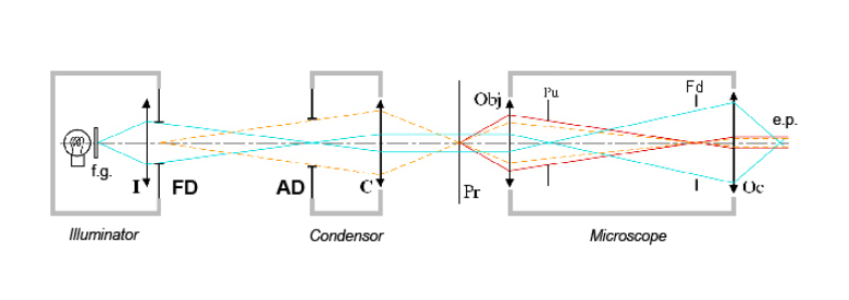
The illumination optic \(I\) is very uniformly illuminated by the lamp and its frosted glass, located a few centimeters behind. The preparation (\(Pr\)), located at the image of lens \(I\) (limited by the field diaphragm \(FD\)) by the condenser \(C\), is therefore uniformly illuminated. However, the frosted glass (whose texture and illumination are less uniform) is imaged at the aperture diaphragm \(AD\). Since this latter element is by construction in the front focal plane of the condenser optics \(C\), the frosted glass is imaged at infinity, as far as possible from the sample.
This configuration has several remarkable properties. Since the aperture diaphragm is located at the condenser focal plane, the system \((AD,C)\) exit pupil is located at infinity (image-space telecentric system). This means that, in addition to being exposed to the same illumination, all the points within the specimen are illuminated by the same light cone, which, considering that the objective is an object-space telecentric system, warrants that all the points in the microscope field of view exhibit the same photometric response. This property is very important in practice and would be very hard to obtain using other configurations... In addition, Köhler illumination allows controlling two important parameters in a totally independent manner:
The illumination field at the specimen, which is controlled by the field diaphragm \(FD\). It is often useful, for parasitic light issues, to limit the illuminated area to the area seen and studied by the microscopist.
The angle of the light cone reaching each point of the preparation, or, in other words, the illumination numerical aperture, controlled by the aperture diaphragm \(AD\). This controls how much light reaches the sample but also, more importantly, how the final image of the observed object is formed; see the sub-section « Illumination coherence and resolution limit », about image formation under partially coherent illumination.
By design, Köhler illumination also ensures that the aperture diaphragm AD is imaged onto the objective pupil \(Pu\). In a similar fashion, the illumination field diaphragm (\(FD\)) is imaged onto the ocular field diaphragm \(Fd\). Consequently, with Köhler illumination, no vignetting is introduced, and the field of view is perfectly uniformly illuminated.
Therefore, to the exception of very low-budget systems, all microscopes now include a Köhler illumination system, which is most of the time pre-aligned, and in particular pre-centered. The optical conjugation field diaphragm/sample (obtained by translation of the condenser) is however left to the user responsibility, therefore allowing the observation of preparations on slides of various thickness.