|
Atomic Force Microscopy (AFM)
The basic principle of the AFM is very similar to a stylus profilometer. Every variation of the surface height influences the force acting on the probe and therefore changes the bending of the cantilever. An image of the observed surface can therefore be created based on the tip deflection information.
AFM technology uses sharp tip (in the range of a few nanometers) at the free end of a cantilever end and detects low forces (a few piconewtons) to provide high resolution information without sample damage. The level of information obtained from AFM
images will depend essentially on the size, shape, and eventually terminal
functionality of the probe tips.
AFM is a powerful analytical tool for characterisation of surfaces in the micro-to nanometer range.
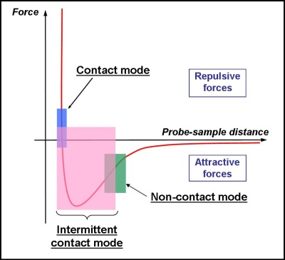
Depending on the type of sample and on the nature of tip-surface interactions, three main operating modes could be used :
- Contact Mode,
- Intermittent Contact Mode (Tapping mode ®)
- and Non-Contact Mode.
In conventional AFM, the bending of the tip is detected by means of a laser beam, which is reflected from the back side of the cantilever into an array of photodiode.
Force, Electric , Magnetic measurements, Nanolithography modes are also possible. |
 |
AFM became a standard microscopy technique for visualizing
and measuring a material’s surface structure in the physical sciences because AFM is capable of measuring nanometer scale images of various nano-object and surfaces with little or no sample preparation.
Physical topography, charge density, magnetic field, and other surface properties can be discerned and measured. AFM differs from other types of non-optical microscopy in that it can image samples under natural conditions - in air or liquids - without keeping the samples under destructive artificial conditions, such as drying, coating with metal, vacuum or freezing.
Materials that can be investigated by AFMs include polymers, metals, crystals and minerals as well as thick and thin film coatings, composites, glasses, synthetic and biological membranes,... . AFM is also a key tool in acquiring kinetic information, and real-time signals of living cells, and is now capable of offering in vivo single-cell diagnostics.
Furthermore, AFM has become recently a powerful tool in biological research. The major advantages of AFM over other tools in biological studies are that no extensive sample preparation is required, and surface features of various biological samples can be examined under physiological conditions.
Besides topographical imaging, the ability of an AFM to quantify the interactions between molecules attached to the tip and molecules at the sample surface opens up an entirely new field for probing the specific interaction forces between surfaces.

Keywords : force, interactions, courbe de force, AFM en contact, tapping mode, pointe fine, mesure en liquide, information cinétique, cellules, forces attractives, forces répulsives, technique d'analyse de matériaux, principe de base, image topographique, image de phase
Laboratoire de Physique de l'Etat Condensé -
Institut de Recherche en Ingénierie Moléculaire et Matériaux Fonctionnels
Faculté du Maine - Université du Maine -
Avenue Olivier Messiaen
72085 Le Mans cedex 09, France
|
|
2010 |
|